


To date, science is known about 280 types of worms that can develop and live in the human body, parasitizing in various organs and tissues.The frequency of human infection depends on the climatic and socio-economic conditions of specific territories (in underdeveloped countries, especially in those located in the tropical and subtropical zone, the level of parasitic infections is much higher than in economically developed states).
Methods of human infection with helminths:
- Biogelmintosis (infection from animals).
- Contagioine helminthoses (transmitted from person to person).
- Geogelmintoses (diseases caused by parasites conducting one of the vital cycles in the Earth).
Factors affecting the manifestations of helminthiases
The method of penetration of the parasite into the body:
- The degree of adaptation of helminth to the human body;
- The density of the population (quantity) of parasitic individuals;
- The worm habitat (tissue parasites live in the thickness of the soft tissues, and the clearing live in the gaps of hollow organs).Some helminths in various phases have an educational and fabric forms.The larval and developing stages of worms, as a rule, cause more pronounced pathological changes.
In the absence of re -infection, the number of adult parasites in the human body does not increase.This feature significantly distinguishes helminthic invasions from diseases caused by bacteria, viruses, fungi and simple organisms.
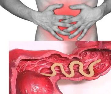
Worms in humans: symptoms
Helminthosis is a disease that is characterized by 2 stages of the course (acute, from two weeks to two months) and chronic (from several months to several years).
Symptoms of acute phase of helminthiasis
The first signs of the disease can manifest itself in different times (most often after 2-3 weeks, with ascaridosis-after 2-3 days, and with phylairiosis, the incubation period can last 6-18 months).
In the acute stage of parasitic invasion, the most characteristic symptom is an allergic reaction (antibodies are generated to antigens of migrating larvae of parasites).Often, itching rashes, prone to the recurrent course, have an increase in the recurrent course, the regional lymph nodes appear on the skin, and the occurrence of generalized or local edema, muscle and articular pains may occur.Also, migrating larvae of the parasite can cause pain in the chest, coughing, suffocation of suffocation, stool disorders, nausea and vomiting.
At the same time, the acute phase of helminthiasis can be accompanied by more serious disorders (severe forms of pneumonia, hepatitis, allergic myocarditis, hepatosterogalia (an increase in the liver and spleen).
In the blood, the amount of eosinophils (eosinophilia) increases and the normal quantitative ratio between protein fractions (dysproteinemia) is disturbed.
Signs of chronic helminthiasis
The symptoms of the chronic phase directly depend on which organ is “populated” with parasites, as well as their size and quantity play an important role.
So, when parasitic in the intestines of single individuals, the disease can occur asymptomatic (with the exception of cases of infection with very large parasites).The characteristic features of the chronic phase of intestinal helminthiasis are dyspeptic disorders.In children, acenoarotic and pain syndrome is more pronounced.With massive invasion ascarides, intestinal obstruction, mechanical jaundice and pancreatitis is possible.
By consuming all the substances necessary for their vital activity from the host’s body, helminths cause digestive disorders, violations of the absorption of vitamins, minerals, carbohydrates, proteins and fats.At the same time, the products of the life of worms inhibit the normal intestinal microflora and reduce the immune forces of the body.
In people suffering from helminthias, due to the weakened immunity and the enhanced process of cellular division (consequences of constant restoration of tissue parasites), the risk of malignant tumors increases significantly.
Types of helminths parasitic in the human body
The causative agents of human helminthiasis are 2 types of worms: round (nematodes) and flat (ribbon and saucers).
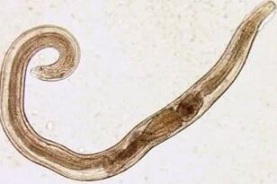
Round worms
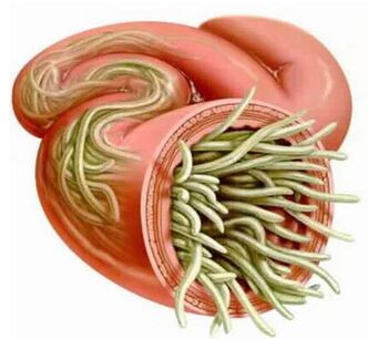
Pinworm
Parasites that are the cause of enterobiosis are small (up to 10mm) thin cavity worms that have grayish-white staining.Infection occurs in an alimentary way (through the mouth).The reason for this is dirty hands.Eggs of the parasite can be in the ground, on the wool of infected animals, unwashed vegetables and fruits, etc. At the same time, with enterobiosis, cases of self -extension (especially in children) that occur as a result of combing areas and subsequent swallows of eggs are not uncommon.The cutting larva develops within two weeks in the digestive tract.Turning into an adult individual, the worm parasitizes in the lower parts of the small and upper parts of the colon.
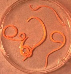
Askarida
Askaride is a large parasite of a spindle-shaped form of red-yellow color, reaching an adult state of 40 cm (females) and 15-25 cm (males).Without a suction cup or other fixing devices, ascaride can independently move towards food masses.Eggs laid down by the female parasite are distinguished with feces.
Acadosis infection occurs in case of swallowing mature eggs along with water or unwashed vegetables and fruits on which there are soil particles.After the eggs penetrate the intestines, ripened larvae come out of them.Then, introducing into the intestinal wall, they reach the heart according to blood flow, and from there they fall into the lungs.Through the pulmonary alveoli, the larva of the ascarida through the respiratory tract penetrates into the oral cavity again.After repeated swallowing, the parasite reaches the small intestine, where it develops into an adult.The worm lives for 12 months, then dies and stands out with feces.In the intestines of one owner can live both one or several hundred individuals.
Vlashev
Vlasov, the causative agent of trichocephalosis, is a white helminth that parasitizes in the initial part of the large intestine and reaches a size of 4-5 cm. The parasite is fed by the blood and tissues of the rectal mucosa.
The sobs -headed eggs laid down by the female on the walls of the intestine come out along with the feces.Their development occurs in the environment (optimally in the soil).Eggs with parasite larvae are penetrated into the body with an alimentary way, through dirty hands, with water or unwashed vegetables and fruits.
Trichinella
The causative agent of trichinellosis is a small round helminth that reaches 2-5 mm in length.Infection occurs when the use of poorly fried meat (pork, bear cubs, wild boar).Penetrating into the intestines, the parasite larva in 3-4 days ripens to the state of the sexually mature individual.The life expectancy of the worm is 40 days, after which the parasite dies.Driving the intestinal wall, larvae penetrate the bloodstream and are carried in all organs of the human body, settling in the muscles.In this case, the respiratory and facial muscles, as well as muscles-ado-bends of the limbs, are most often affected.
In the first days after invasion, patients complain of abdominal pain.Then, after about 2 weeks, the body temperature rises to 39-40 s, itchy rashes appear on the skin, muscle pain develops, and the face swells.In this period, in the case of massive infection, there is a significant risk of death.After about a month, recovery occurs.The parasite is encapsulated in a spiral form, after which it dies within two years.
Ankylostoma and non -core
These two parasites are similar to each other by biological characteristics, as well as in the caused diseases.With this, it is customary to unite them under the general name (ankylostoma).Worms reaching lengths 10-15 mm are parasitized in 12-P.gut.It should be noted that this is one of the most common, but at the same time, rarely identified parasites.Worms of worms penetrate the human body through the skin when in contact with infected soil.Further, getting into the bloodstream, they, just like ascarides, migrate to the lungs, and then, through the bronchi, along with the hopping sputum - to the digestive tract.Anquilostoma parasitizes in the intestines, attaching to the intestinal wall.A parasite that eats exclusively with blood bites the blood vessels piercing the mucous membrane, injecting an anti -impoverished component there.During the day, an adult can absorb 0.05-0.35 ml of blood on average.Therefore, the most characteristic symptom of this helminthiasis is iron deficiency anemia, as well as a change in the ratio of protein fractions (dysproteinemia).
Flat worms
Wide tape
This is one of the largest helminths reaching a length of 10-20 meters.The disease caused by this parasite is called dipillobotriosis.The worm development cycle begins with freshwater fish or crustaceans.The larva enters the human body, which is the final owner of a wide ribbon with caviar or infected fish fillet.Reaching the small intestine, the parasite is attached to its wall and for 20-25 days grows to a sexually mature individual.
Hepatic bacon
A parasite causing opisthorchiasis is a flat worm reaching a length of 7-20 mm.It should be noted that more than 50% of cases of infection with a liver saucer (it is also called a cat biconomater) falls on the inhabitants of Russia.The parasite larvae begin to develop after the eggs fall into fresh water (from the snails swallowed).Then they penetrate the body of the fish (carp, crucian carp, bream, roach).Human infection occurs when you eat infected fish meat that has not undergone sufficient heat treatment.The larva of the hepatic bomb from the small intestine penetrates the biliary ducts and into the gall bladder, fixing there with two suction cups.
Bull and pork
These parasites almost identical in structure reach a length of 5-6 meters.Infection with tusiarinhosis and tusiasis occurs due to the use of cattle or pork meat infected with Finns (one of the intermediate forms of helminthiasis).Visible Finns, presented in the form of whitish bubbles, reaching 0.5 cm in size, are attached to the wall of the small intestines of a person and in 3 months turn into an adult individual.The strip parasite, consisting of more than 2000 segments, is constantly growing.At the same time, the end segments containing eggs come off and independently move along the colon to the anal opening, and then crawl out of the anal, or stand out into the external environment along with feces.The most characteristic symptoms of helminthiasis are a violation of the digestive tract.
Echinococcus
For this parasite, a person is an intermediate host.The worm parasitizes in the human body in the form of Finns.The final owner of Echinococcus is a wolf, a dog or a cat.Infection occurs in an alimentary way in contact with animals and environmental objects, with the handful of echinococcus eggs.After getting into the intestines, oncospheres (six -black larvae) develop from them.From the intestine they penetrate the bloodstream and are carried throughout the body.
Alveokokk
This parasite, which is considered a variety of echinococcus, is the cause of one of the most dangerous helminthiases (alveococcosis), which is similar in severity with cirrhosis and liver cancer.Infection occurs with the penetration of oncosphere (eggs with ripened larvae) into the intestines.There, the embryo comes out of the egg and, introducing into the intestinal walls, penetrates into the bloodstream.Further, with a blood flow, the parasite spreads through all tissues and organs of the body (most often localized in the liver).It is there that the main stage of development begins at the larvae (a multi -chamber bubble, laurelocyst) is formed).Each chamber contains an embryo head of a parasite, which continues to gradually develop.Lavrocists are very aggressive formations that constantly grow due to increasing bubbles, as well as have the ability to germinate into the liver, like cancer metastases.
Diagnosis of helminthiasis
Diagnosis of helminthic invasions includes the following events:
- A thorough collection of an anamnesis that helps to find out the possible causes of infection;
- Laboratory studies of feces, blood, the contents of 12P intestines, rectal and perianal mucus, muscle tissue, pulmonary sputum, bile.During the analysis, eggs, segments or fragments of parasites can be detected.At the same time, the increased content of eosinophils in the blood is also a signal about the presence of helminthiasis.
- In the diagnosis of diseases caused by the larval stages or tissue parasites, serological studies (ELISU, RSC, indirect agglutination, immunofluorescence analysis, etc.) are carried out.
- To identify helminths that affect the liver tissue, ultrasound, CT and endoscopic studies are prescribed.
Worms in humans: treatment
In the acute phase of a parasitic infection, the patient is prescribed detoxification and desensitizing therapy.In a severe course of the disease (liver trematodosis, trichinellosis), glucocorticoids are used according to medical indications.
As drugs of specific therapy, taking into account the nature of the pathogen, special anthelmintic chemotherapeutic agents are prescribed.
In parallel, the patient is recommended to take antihistamines and enterosorbents.The final stage of treatment includes the use of probiotics that normalize the intestinal microflora.
A special sparing diet is also prescribed (food should be easily digestible and contain little fat).
During the period of anthelmintic therapy, the patient needs strict adherence to personal hygiene (in order to avoid re -infection).At the same time, with many helminthiasis, all family members and persons who are infected in constant contact should undergo treatment.